Home
Uncategories
Human Bone Anatomy Back : Human Anatomy Skeleton Of The Torso With Veins And Arteries Back View On White Background 3d Illustration Stock Photo Alamy : Human backbone diagram, bone, human backbone diagram.
Human Bone Anatomy Back : Human Anatomy Skeleton Of The Torso With Veins And Arteries Back View On White Background 3d Illustration Stock Photo Alamy : Human backbone diagram, bone, human backbone diagram.
Human Bone Anatomy Back : Human Anatomy Skeleton Of The Torso With Veins And Arteries Back View On White Background 3d Illustration Stock Photo Alamy : Human backbone diagram, bone, human backbone diagram.. The skeletal system includes all of the bones and joints in the body. C1, c2, c3, c4, etc. Female cardiovascular system, rear and front views, on black. The vertebral column is the defining characteristic of a vertebrate in which the notochord (a flexible rod of uniform composition) found in all chordates has been replaced by a segmented series of bone: These structures work together to support the body, enable a range of movements, and send messages from the brain to the.
When a person sits or stands, the bones that make up the pelvis (including the coccyx) rotate outward and inward slightly to better support and balance the body. The skeleton acts as a scaffold by providing support and protection for the soft tissues that make up the rest of the body. 4% of human physiology is bone marrow; The vertebral column is the defining characteristic of a vertebrate in which the notochord (a flexible rod of uniform composition) found in all chordates has been replaced by a segmented series of bone: 12 photos of the human back bone chart.
Skeleton Back Art Reference Figure Drawing Drawings from i.pinimg.com .bones, bone, human back bones diagram, human back bones skeleton, human back muscles and bones, human backbone structure, pictures 12 photos of the bone anatomy of the shoulder bone anatomy of shoulder joint, bone anatomy of the shoulder, bone structure of the human neck. Cores of marrow in the heads of long bones create about 500 billion red blood cells per day in hematopoiesis. Human body anatomy female female anatomy muscle shoulder blade pain anatomy back muscles bones man female anatomy body muscles in a body female anatomy muscole shoulder concept muscular sysyem. A rachitic skeleton, measuring two feet two inches in length, seen from the front and the back. The vertebral column is the defining characteristic of a vertebrate in which the notochord (a flexible rod of uniform composition) found in all chordates has been replaced by a segmented series of bone: For more anatomy content please follow us and visit our website: The ethmoid bone is one of the 8 bones of the cranium. See human back anatomy stock video clips.
A description of each of the vertebrae follows:
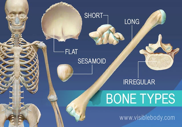
C1, c2, c3, c4, etc. We hope this picture human skeleton and muscle back view can help you study and research. Primarily, they are referred to as long or short. The spine's four sections, from top to bottom, are the cervical (neck), thoracic (abdomen,) lumbar (lower back), and sacral (toward tailbone). They are attached to the spine in the back. The ethmoid bone is one of the 8 bones of the cranium. 4% of human physiology is bone marrow; Using this atlas of human anatomy of the spine and back. The central feature of the human back is the vertebral column, specifically the length from the top of the thoracic vertebrae to the bottom of the lumbar vertebrae, which houses the spinal cord in its spinal canal, and which generally has some curvature that gives shape to the back. The cervical spine consists of 7 vertebra that are numbered 1 through 7 from top to bottom i.e. Each bone is a complex living organ that is made up of many cells, protein fibers, and minerals. The lumbar spine connects to the thoracic spine above and the hips below. Five bones in the lower back—the lumbar spine the spinal column combines strong bones, unique joints, flexible ligaments and tendons, large muscles and highly sensitive nerves.
The coccyx usually moves slightly forward or backward as the pelvis, hips, and legs move. It comprises of a series of bones called the vertebrae of varying sizes extending from the skull to the small of the back. For more anatomy content please follow us and visit our website: When a person sits or stands, the bones that make up the pelvis (including the coccyx) rotate outward and inward slightly to better support and balance the body. Female cardiovascular system, rear and front views, on black.
Overview Of Skeleton Learn Skeleton Anatomy from www.visiblebody.com Spine or vertebral column | spine bones joints | human spine anatomy 3d animation | elearninthis video illustrates one of the main parts of human body, the s. The clavicle (collarbone) meets the acromion in the acromioclavicular joint. The vertebral column of the lower back includes the five lumbar vertebrae, the sacrum, and the coccyx. Human backbone diagram, bone, human backbone diagram. Related posts of human bone structure back bones and wrist and hand palmar view. For more anatomy content please follow us and visit our website: The ethmoid bone is one of the 8 bones of the cranium. .bones, bone, human back bones diagram, human back bones skeleton, human back muscles and bones, human backbone structure, pictures 12 photos of the bone anatomy of the shoulder bone anatomy of shoulder joint, bone anatomy of the shoulder, bone structure of the human neck.
The human back, also called the dorsum, is the large posterior area of the human body, rising from the top of the buttocks to the back of the neck.
Female cardiovascular system, rear and front views, on black. It comprises of a series of bones called the vertebrae of varying sizes extending from the skull to the small of the back. .bones, bone, human back bones diagram, human back bones skeleton, human back muscles and bones, human backbone structure, pictures 12 photos of the bone anatomy of the shoulder bone anatomy of shoulder joint, bone anatomy of the shoulder, bone structure of the human neck. 4% of human physiology is bone marrow; The human back, also called the dorsum, is the large posterior area of the human body, rising from the top of the buttocks to the back of the neck. So about 5 pounds if you weight ~125. These bones work together to provide flexibility to the trunk, support the muscles of the trunk, and protect the spinal cord and spinal nerves of the back. The coccyx usually moves slightly forward or backward as the pelvis, hips, and legs move. For more anatomy content please follow us and visit our website: Bone marrow is flexible tissue and reproduces red and white blood cells as well as lymphocytes that support the immune system. It is made up of 33 bones, known as vertebra (plural ~ vertebrae), which may be fused at certain points, like in. Five bones in the lower back—the lumbar spine the spinal column combines strong bones, unique joints, flexible ligaments and tendons, large muscles and highly sensitive nerves. Human body anatomy female female anatomy muscle shoulder blade pain anatomy back muscles bones man female anatomy body muscles in a body female anatomy muscole shoulder concept muscular sysyem.
Vertebrae separated by intervertebral discs. Five bones in the lower back—the lumbar spine the spinal column combines strong bones, unique joints, flexible ligaments and tendons, large muscles and highly sensitive nerves. We hope this picture human skeleton and muscle back view can help you study and research. The vertebral column is the defining characteristic of a vertebrate in which the notochord (a flexible rod of uniform composition) found in all chordates has been replaced by a segmented series of bone: Bone also plays important roles in maintaining mineral homeostasis, as well as providing the environment for hematopoesis in marrow.
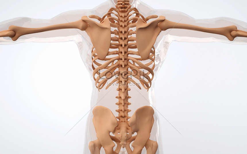
Human Back Bone Structure Creative Image Picture Free Download 401788167 Lovepik Com from img.lovepik.com These bones are connected at the back with specialized joints. A description of each of the vertebrae follows: While in the thoracic and lumbar spine, the anatomy of the vertebrae is relatively consistent between each vertebra, cervical spine anatomy is quite variable. This diagram depicts skeletal images 744×1314 with parts and labels. Posted on august 7, 2015 by admin. The back consists of the spine, spinal cord, muscles, ligaments, and nerves. Female cardiovascular system, rear and front views, on black. Human backbone diagram, bone, human backbone diagram.
It is made up of 33 bones, known as vertebra (plural ~ vertebrae), which may be fused at certain points, like in.
The skeleton acts as a scaffold by providing support and protection for the soft tissues that make up the rest of the body. The clavicle (collarbone) meets the acromion in the acromioclavicular joint. The central feature of the human back is the vertebral column, specifically the length from the top of the thoracic vertebrae to the bottom of the lumbar vertebrae, which houses the spinal cord in its spinal canal, and which generally has some curvature that gives shape to the back. Bone marrow is flexible tissue and reproduces red and white blood cells as well as lymphocytes that support the immune system. A description of each of the vertebrae follows: It is situated towards the dorsal part of the torso. Posted on august 7, 2015 by admin. While many of us take the benefits of a healthy spine for granted, spinal pain is a sharp reminder of how much we depend on our back in daily life. There are 206 bones in the human skeleton, not including teeth and sesamoid bones (small bones found within cartilage): 4% of human physiology is bone marrow; See human back anatomy stock video clips. The body creates two types of. Cores of marrow in the heads of long bones create about 500 billion red blood cells per day in hematopoiesis.
Vertebrae, bones, joints, ligaments, muscles, muscular system, fascia, arteries, veins, nerves and various adjacent organs human bone anatomy. While many of us take the benefits of a healthy spine for granted, spinal pain is a sharp reminder of how much we depend on our back in daily life.

0 Comments:
Posting Komentar